

Medical Oncologist
Most of the fear is about the room and the machine, not the result. Here is the whole visit, step by step — including why it presses, and how long that part really lasts.
It is an X-ray of the breast tissue, taken between two flat plates. Nothing enters the body. Nothing is injected, and there is no needle.
Two pictures are usually taken of each side, from different directions, so that overlapping tissue does not hide anything.
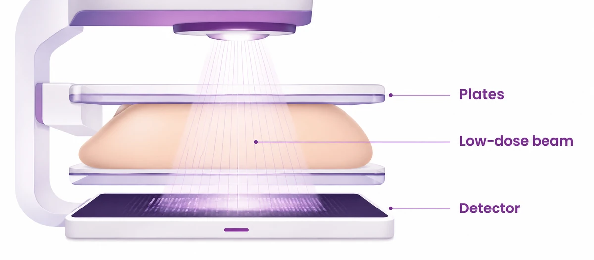
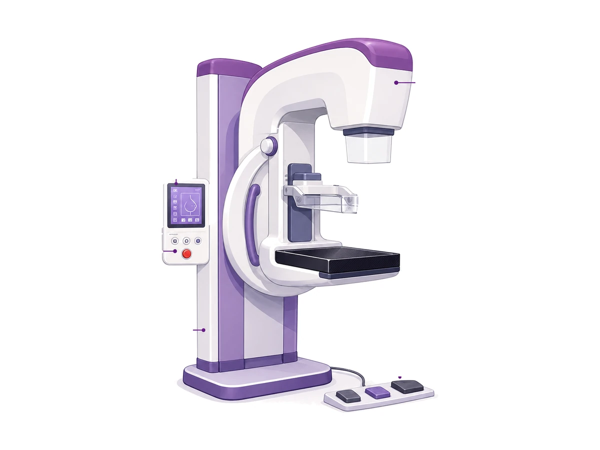
Usually the same machine — in 3D the tube sweeps in a small arc and builds the picture in thin slices rather than one flat image. What happens to you is almost identical: same position, same compression, a few seconds longer.
| Comparison | 2D | 3DThe better picture |
|---|---|---|
| Cancers found | The baseline every study compares against. | Roughly one or two more per 1,000 women scanned. |
| Being called back | Around one woman in ten is asked to return for another look. | Fewer in most studies — and when you are called back, it is more often for something real. |
| Dense tissue | Hardest to read. Overlapping layers both hide and mimic. | The biggest gain is here, because separating the layers is exactly what dense tissue needs. |
| Radiation | One exposure per view. | Essentially the same dose. The flat image is generated from the same set of pictures. |
| In the room | A few seconds per view. | A few seconds longer. Nothing else differs. |
Nearly all the worry before a first mammogram is not knowing the sequence. Here it is.
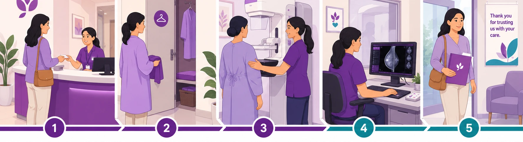
You change in a private cubicle and only the top half is uncovered, with a female radiographer.
The whole appointment is short. The part where the plates press lasts seconds per picture.
A radiologist reads the images afterwards. Ask when and how you will be told before you leave.
Say clearly when you call that you have noticed something, so you are booked for the right kind of appointment. Most breast lumps turn out not to be cancer. But the way to find that out is to have it looked at — not to wait for the next screening round.
Or call 1800 202 8726 — toll free, no obligation.
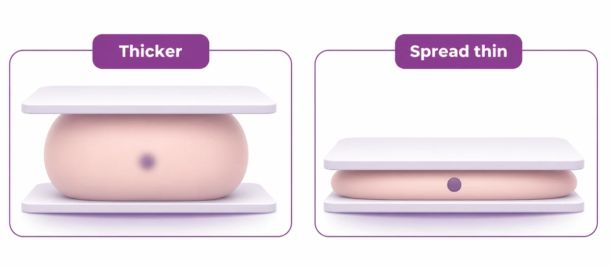
The plates press firmly, and this is the part people dread. For most it is uncomfortable for a few seconds rather than painful.
Spreading the tissue thinner is what lets a small thing show up. It is like flattening dough to see what is mixed into it — you cannot see through a thick lump of it.
It lasts seconds, not minutes. And the radiographer can ease it if you ask.
Talk to our team
Call the helpline or leave your details, and someone will help you arrange a consultation at the CION centre nearest you. One helpline serves every CION centre.
Being sent for an ultrasound as well does not mean the mammogram failed. They answer different questions.
Good at finding tiny specks of calcium and subtle changes in structure — often before anything can be felt.
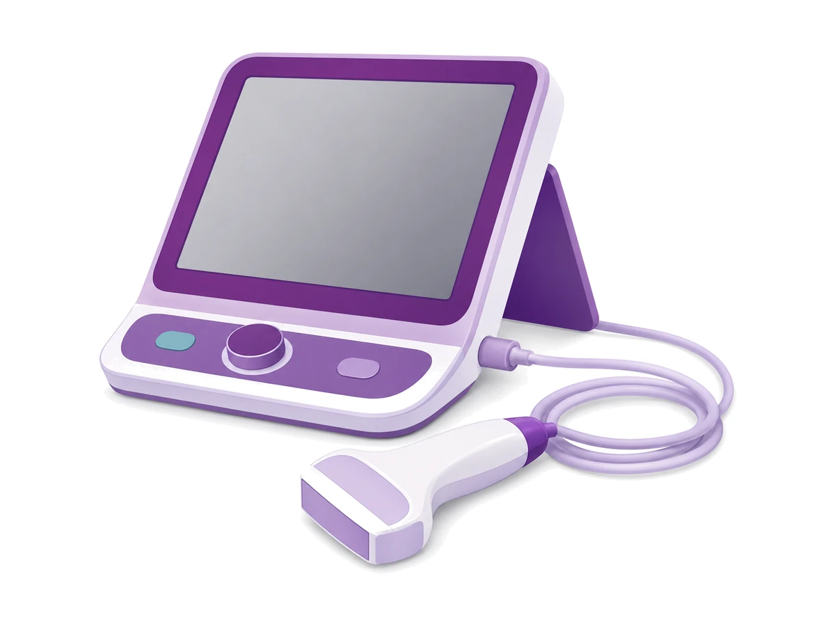
Good at telling whether something is solid or just fluid. Often added when the tissue is dense, or to look closely at one spot.
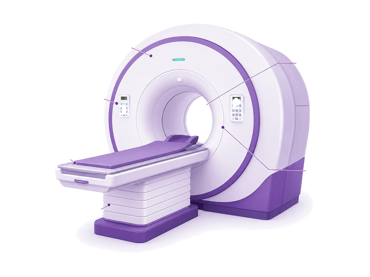
Used in particular situations rather than routinely — for example when someone is known to be at higher risk.
Neither is better than the other. An added scan is thoroughness, not a sign that something was found. If your report mentions dense tissue, that is explained here →
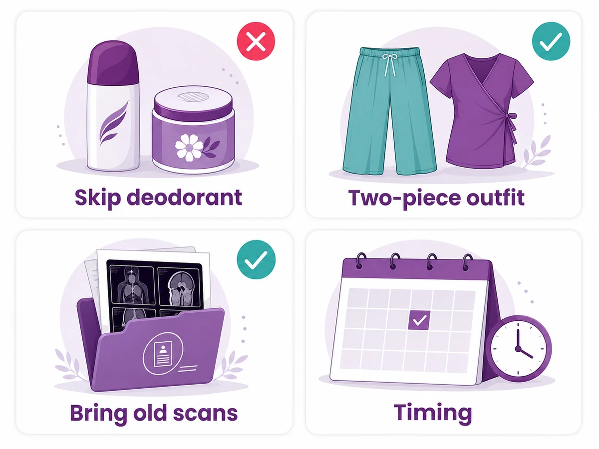
Tell them when you book if you have implants. A different technique is used to get a clear picture around them, and the appointment takes a little longer. It is routine — they just need to know in advance.
Tap any of these. Each one keeps women away from a test that is quick and, for most, unremarkable.
Worried about something else? Tell us what is on your mind — no obligation to book.
Ask us directlyThe dose used is very small — far less than most people picture when they hear the word X-ray. Set against that is a real benefit: finding a change early, when there are more options and usually less treatment.
What to do: if you are pregnant or might be, say so before the appointment. That does change what is recommended.
General information, not advice about your case. If you have noticed a change in your breast, do not wait for a screening appointment — call 1800 202 8726.
Still unsure whether to book? Call and describe your situation. We will tell you honestly whether it is worth booking now or waiting.
Talk it through firstTwo facts, and they are connected.
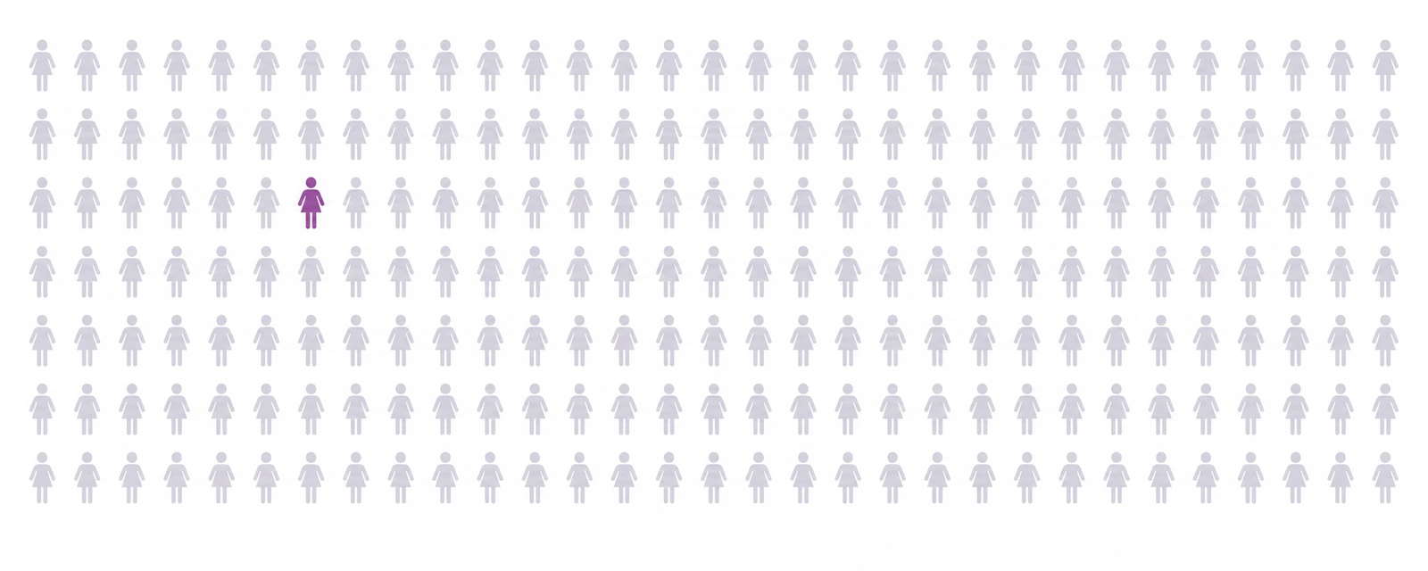
Only about one woman in 250 in Telangana has ever had her breasts checked by a doctor.
And more than half of breast cancers in India are found only after they have already grown, or reached the nearby lymph nodes.
Those two facts are connected. Being in the majority here does not make it safe. It is the reason so many are found late, when more treatment is needed than would have been.
Short explainers covering the same ground as this page. Nothing loads or plays until you press it.
Awaiting URL
Awaiting URL
Trained at AIIMS, Tata Memorial, and leading international centres. Combined 150+ years of experience. Every complex case is reviewed by 3+ of them — together.

MBBS(Gold Medal), DNB(General Medicine), DM(Medical Oncology)(Gold Medal)

MBBS, MD(General Medicine), DM(Medical Oncology)(Adyar,Chennai), ECMO, MRCP SCE(UK)

MBBS, MD (General Medicine), DrNB (Medical Oncology), ECMO, MRCP SCE (Medical Oncology) (UK)



MBBS (AIIMS), MS (Surgery) (AIIMS), DNB (Surgical Oncology), MRCS (Edinburgh)



MBBS, MS(General Surgery), M.Ch(Surgical Oncology), FMAS, FARIS(Ongoing)






MBBS, MS (General Surgery), DrNB (Surgical Oncology), FALS Oncology

Want a specific doctor for your case? Mention them when booking.
Book Free ConsultationShare your name and number — we'll call you back within 30 minutes to schedule your consultation.
Where to find us
Addressed by landmark, because that is how this city navigates. Each centre also names the areas it serves, so you can place it without a map. Consultation and day-care Chemotherapy run at every one of them.
Talk to our team
Call the helpline or leave your details, and someone will help you arrange a consultation at the CION centre nearest you. One helpline serves every CION centre.
The six we hear most often before a mammogram. Tap any question to open the answer.
The plates press firmly, and that is the part people dread. But it lasts seconds rather than minutes, and the radiographer can ease it if you ask. The pressing is not incidental — spreading the tissue thinner is what lets a small change show up at all. If you have found it very painful before, say so when you book. Timing the appointment for a more comfortable point in the month often helps. So does asking the radiographer to go more slowly. Fear of pain is one of the commonest reasons women skip this test, and it is usually worse in the imagining.
This worry stops a lot of people going, and it is understandable — the word X-ray sounds alarming. The dose used is very small, far less than most people picture. Set against that is a real benefit: finding a change early, when there are more options and less treatment is usually needed. Your radiographer can tell you exactly what dose the machine at your centre uses if you would like to know. If you are pregnant or think you might be, tell them before the appointment, because that does change what is recommended.
This is probably the most damaging myth about breast cancer, because it feels so reasonable. A strong family history does raise risk, and it does change what is recommended. But the large majority of women who develop breast cancer have no family history at all. Having none is not protection and is not a reason to skip a check. If you do have close relatives affected, mention it. It may mean starting earlier or adding a different test. That is a conversation worth having rather than assuming.
Being called back is frightening and it is very often nothing. The commonest reason is simply that the images were not clear enough to be sure. Overlapping tissue, a slightly off position, or no earlier scan to compare against. That is recorded as an incomplete result rather than a finding. What happens next is usually more images, sometimes an ultrasound. Go promptly so you get an actual answer instead of sitting with an unfinished one, and take any previous scans with you.
In 2D the tube takes one picture per view, so everything in the breast is layered into a single flat image. In 3D it sweeps in a small arc and takes several, which are rebuilt into thin slices. That helps most where the tissue is dense, because overlapping layers are exactly what makes a 2D picture hard to read. It is usually the same machine doing both. The experience is almost identical, just a few seconds longer, and it does not mean two exposures because the flat image is generated from the same set of pictures. Whether it is worth the extra depends on your case. And if your centre still uses a film machine, where the picture ends up on a sheet rather than a screen, ask whether you can also have the images on a CD.
The two tests answer different questions. A mammogram is good at picking up tiny specks of calcium and subtle changes in structure. An ultrasound is good at telling whether something is solid or just fluid. An ultrasound is often added when the tissue is dense, or to look more closely at one specific area. Being sent for one is thoroughness rather than bad news, and it is very common. Neither test is better than the other; they are simply used for different things.
A screening mammogram is for when nothing has been noticed. If you have felt a lump or seen a change, that is a different situation. It needs a proper assessment, not a routine appointment slot. Say clearly when you call that you have felt something, so you are booked for the right kind of visit. Most breast lumps turn out not to be cancer. But the way to find that out is to have it looked at, rather than waiting for the next screening round.
If a scan does lead further, these run alongside treatment at CION rather than as afterthoughts.
Appetite, weight and strength through treatment.
Support through uncertainty, for patient and family.
Hereditary risk for you and close relatives.
Comfort and symptom support at any stage.
Dedicated pain control as part of the plan.
Shoulder mobility and lymphoedema care.
Tell us if you have noticed anything, so we book you for the right kind of visit. Bring any previous scans, films or a CD from another centre. Comparing against an older picture often settles a question on the spot.
Toll free 1800 202 8726 · Advancing Care. Passionately.