


Medical Oncologist
Your mammogram report ends with a number nobody explained. Here is what each category means, what happens next — and why category 4 is not stage 4.
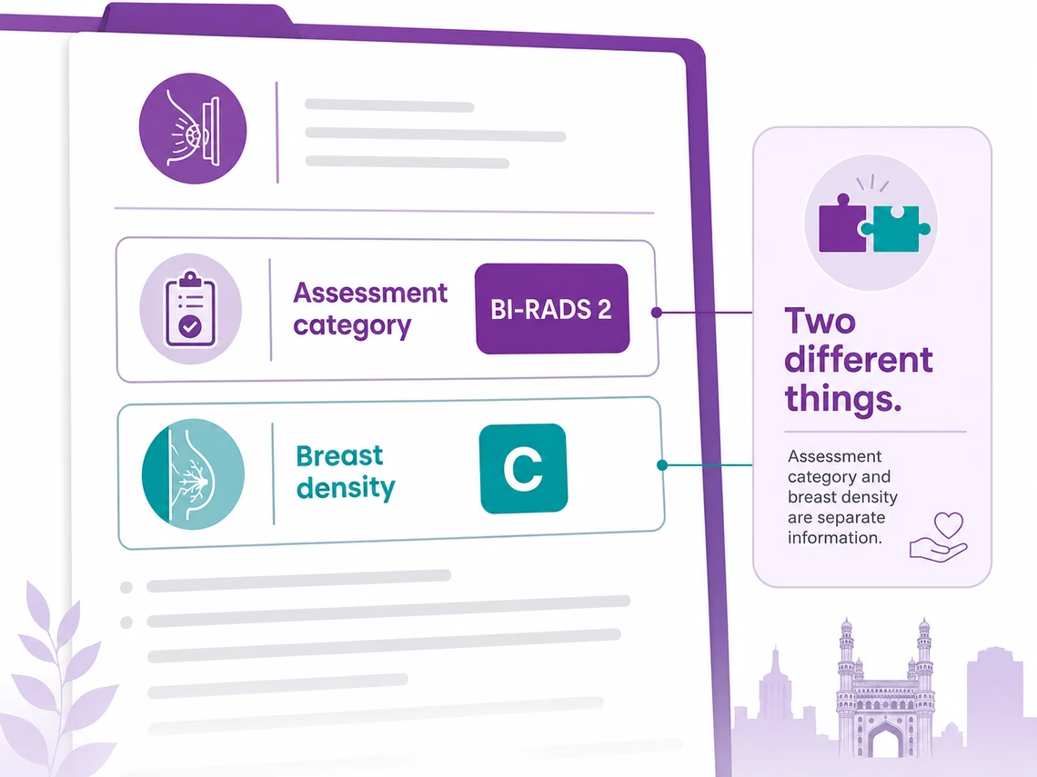
A mammogram report carries two separate graded results, and they are routinely confused with each other.
The category is the radiologist telling you how confident she is — like a weather forecast giving a chance of rain. A forecast is not the weather, and a category is not a diagnosis.
Most findings placed in category 4 turn out not to be cancer. The number describes the picture, not you.
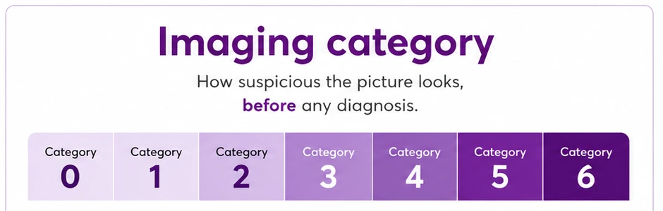
The categories are a scale of concern, not a scale of severity. Each one carries a different next step. Find yours.
 Category 0
More images needed. This is not a finding.
 Category 1
Nothing found. Back to routine screening.
 Category 2
Clearly not cancer. Back to routine screening.
 Category 3
Short-interval follow-up to confirm it does not change.
 Category 5
Biopsy strongly advised. Planning often begins in parallel.
 Category 6
A biopsy has already proven cancer. The scan is for planning.
Not sure which category you have been given? Send us your report. A specialist will tell you what it says and whether anything needs doing now.
Get my scan report explainedTap the number on your report to see what it means, how worried to be, and what usually happens next.
If your report shows a letter A to D as well, that is breast density — a separate thing. Explained below ↓
Very likely harmless. The radiologist is not worried, but wants to confirm the finding does not change over a short interval.
What usually happens: a repeat scan after a short interval rather than a biopsy. If nothing changes, you return to routine screening.
General explanations to help you follow your own report — not a diagnosis, and no substitute for a specialist reading your images. No category is a confirmed cancer; only a biopsy can decide that. Call 1800 202 8726 if your report does not match anything here.
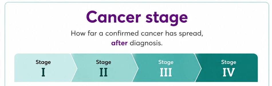
This is the single most distressing misreading of a mammogram report, and it happens constantly. They are not the same scale, and not even the same subject.
Both use small numbers, and that is the only thing they share. A hotel star rating and a room number use the same digits and mean completely unrelated things.
If you have been given a category 4, you do not have a stage. You have a recommendation for a biopsy. How staging actually works →
Talk to our team
Call the helpline or leave your details, and someone will help you arrange a consultation at the CION centre nearest you. One helpline serves every CION centre.
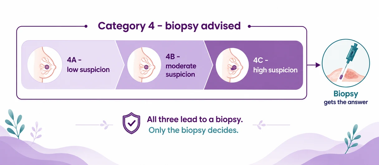
If your report says 4A, that is the mildest of the three — and most people reading a bare "4" assume the worst available meaning of the number.
The letters are not degrees of how bad it is. They describe how strongly the picture argues for a biopsy. All three end in the same place — a biopsy, which is the only thing that actually decides.
Even 4C is not a diagnosis. It is a strong reason to find out.
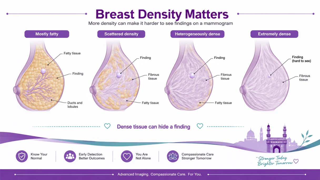
Density is reported on every mammogram, as a letter from A to D. It is not a disease and not a diagnosis — it describes how much dense tissue you naturally have.
It matters for one practical reason, and it explains why an ultrasound is often added.
On a mammogram, dense tissue is white and a lump is also white. It is like trying to spot a white cricket ball on a white bedsheet instead of on a green field.
Dense breasts are a normal finding, and they are more common in younger women. If your report mentions density, it is telling the next doctor how easy or hard the picture was to read.
The descriptive words on a mammogram report are the most opaque part of it. Tap any you can see.
A three-dimensional lump the radiologist can see on more than one view. The word says nothing about whether it is harmless.
Why it matters: what follows it on the report — the shape and the margin — is what actually moves the category up or down.
General explanations to help you follow your own report — not a diagnosis. If a word on your page isn’t here, call 1800 202 8726.
Ask for the films or the CD. Any second reading needs the images themselves, not the printed summary.
Including the letter if there is one. 4A and 4C lead to the same next step but very different levels of concern.
Both mean a biopsy is advised. Booking it is the fastest route out of uncertainty.
Been advised a biopsy? How to read the report you will get →
Send us the report and the images. A specialist will tell you whether the recommended next step is the one we would advise.
Or call 1800 202 8726 — toll free, no obligation.
Trained at AIIMS, Tata Memorial, and leading international centres. Combined 150+ years of experience. Every complex case is reviewed by 3+ of them — together.

MBBS(Gold Medal), DNB(General Medicine), DM(Medical Oncology)(Gold Medal)

MBBS, MD(General Medicine), DM(Medical Oncology)(Adyar,Chennai), ECMO, MRCP SCE(UK)

MBBS, MD (General Medicine), DrNB (Medical Oncology), ECMO, MRCP SCE (Medical Oncology) (UK)



MBBS (AIIMS), MS (Surgery) (AIIMS), DNB (Surgical Oncology), MRCS (Edinburgh)



MBBS, MS(General Surgery), M.Ch(Surgical Oncology), FMAS, FARIS(Ongoing)






MBBS, MS (General Surgery), DrNB (Surgical Oncology), FALS Oncology

Want a specific doctor for your case? Mention them when booking.
Book Free ConsultationShare your name and number — we'll call you back within 30 minutes to schedule your consultation.
Where to find us
Addressed by landmark, because that is how this city navigates. Each centre also names the areas it serves, so you can place it without a map. Consultation and day-care Chemotherapy run at every one of them.
Talk to our team
Call the helpline or leave your details, and someone will help you arrange a consultation at the CION centre nearest you. One helpline serves every CION centre.
The six we hear most often after a mammogram result. Tap any question to open the answer.
Category 4 says the picture is not clear-cut, so tissue is needed to settle it. Most findings placed in category 4 turn out not to be cancer, and that is true even of the higher sub-levels. The category describes how strongly the image argues for a biopsy, not what the biopsy will show. It is also worth checking whether your report specifies 4A, 4B or 4C, because 4A carries considerably less suspicion than 4C while leading to the same next step. Nothing in a category is a diagnosis. Only the biopsy decides, and booking it is the fastest way out of the uncertainty.
Category 3 means the radiologist thinks a finding is very likely harmless but wants to confirm it does not change. The standard response is a repeat scan after a short interval rather than a biopsy. That is a defined pathway with decision points built into it, not a decision to leave things alone: if the repeat scan shows no change you return to routine screening, and if it changes you move to a biopsy. It is not a compromise or a cost decision. If waiting makes you uneasy, it is entirely reasonable to ask for a second reading of the images in the meantime.
Category 0 is not a finding and not a low score on a scale of badness. It means the images available were not enough to reach a conclusion, so more are needed — extra mammogram views, an ultrasound, or comparison with a previous scan. It happens often, particularly on a first mammogram where there is nothing earlier to compare against, and in women with dense tissue. Being recalled after a category 0 is not evidence that something was seen. Book the additional imaging promptly so you get an actual answer rather than sitting with an unfinished one.
Breast density describes how much working tissue you have compared with fat, graded A to D. It is a normal variation, it is more common in younger women, and it is not a diagnosis. It matters for one practical reason: on a mammogram, dense tissue appears white and so does a lump, so dense tissue can hide a small finding. That is why an ultrasound is often added when density is high. If your report mentions density, read it as a note to the next doctor about how easy or hard the picture was to read, not as a warning about you.
Calcifications are tiny deposits of calcium and they appear on a very large share of mammogram reports. The great majority are harmless and need nothing done. What the radiologist assesses is the pattern rather than the presence: specks that are larger, rounder and widely scattered are ordinary, while very fine specks grouped tightly in an irregular cluster earn a closer look. Even then, most clustered calcifications still turn out benign after a biopsy. They are, however, the commonest reason a category 4 is given when nothing at all can be felt.
A second reading is routine practice, not a challenge to the first radiologist. It is most worth doing when you have been given a category 3 or 4, when the report and your own examination do not seem to agree, or when you are being asked to wait and are not comfortable with it. Bring the actual images rather than only the printed report, because a second reading needs what the first radiologist saw. Ask for the films or the CD from wherever the scan was done, and keep them with your other records.
Want your images read again? Bring the films or the CD. We will tell you honestly whether we read them the same way.
Request a second readingIf your scan does lead further, these run alongside treatment at CION rather than as afterthoughts.
Appetite, weight and strength through treatment.
Support through uncertainty, for patient and family.
Hereditary risk for you and close relatives.
Comfort and symptom support at any stage.
Dedicated pain control as part of the plan.
Shoulder mobility and lymphoedema care.
A specialist will read your report and your images, explain the category in plain terms, and tell you whether the recommended next step is the one we would advise. Bring the films or the CD if you have them.
Toll free 1800 202 8726 · Advancing Care. Passionately.