

Medical Oncologist
You are holding a page full of words nobody has explained to you. Here is what each line means — and which two actually change your treatment.
Every breast biopsy report answers the same six questions. Two of them move your treatment more than the rest.
The report is a report card on the tumour, not on you. Like a soil test before you decide which crop to plant — the test doesn’t change the field, it decides the plan. It describes the tumour as it is today; it does not forecast the person, and no single line on it is a verdict.
Laboratories format reports differently, but almost all carry these blocks in roughly this order. Find each one on your own page.
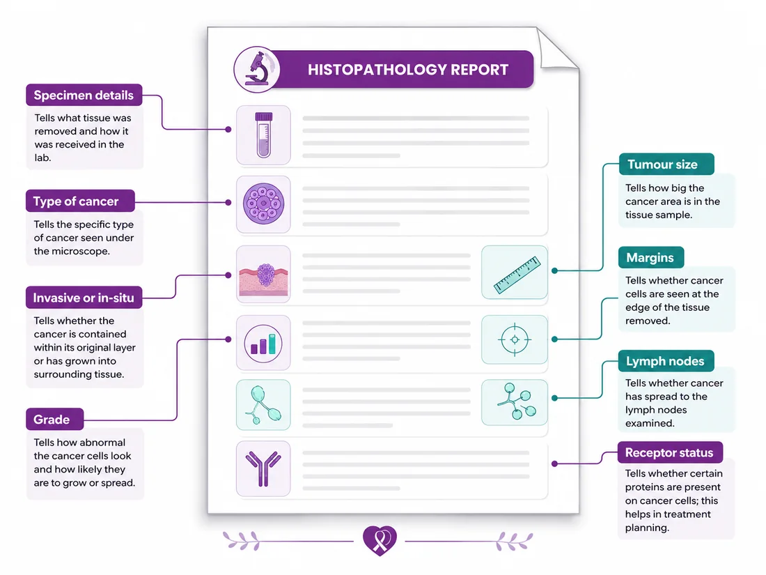
Which breast, which side, how the sample was taken, when. Check it — a wrong side does occasionally appear, and it is easier to correct now.
Where the cells began. Tells you the starting point, not how serious it is. See the diagram ↓
Still inside the duct, or crossed its wall. Decides whether lymph nodes need assessing. Explained below ↓
How closely cells still resemble normal tissue. Not the same as stage. See the diagram ↓
Measured on the tissue removed. Can change once more tissue is examined.
The rim of healthy tissue around what was removed. Usually appears only after surgery. See the diagram ↓
Whether underarm nodes contained cancer cells. How nodes affect stage →
What fuels the cancer, and which treatments can work against it. Often issued days later. Why ↓
Can't match your report to this? Reports vary between laboratories. Send us yours and an oncologist will go through it line by line — no obligation.
Get my report explainedTap any word you can see on your page. Covers the terms that appear on most breast pathology reports.
The cancer cells have crossed out of the duct or lobule where they started, into the surrounding breast tissue.
Why it matters: once cancer is invasive, the lymph nodes are assessed and the treatment discussion widens.
General explanations to help you follow your own report — not a diagnosis, and no substitute for your oncologist reading your case. If a word on your page isn't here, call 1800 202 8726.
Picture a water pipe running through a wall. In situ is water still inside the pipe. Invasive is water that has come through the pipe wall into the wall of the house. But don’t hear “in situ” as harmless — it is a cancer that has not yet crossed the wall, and it still needs treatment. People who read it as nothing sometimes delay care.
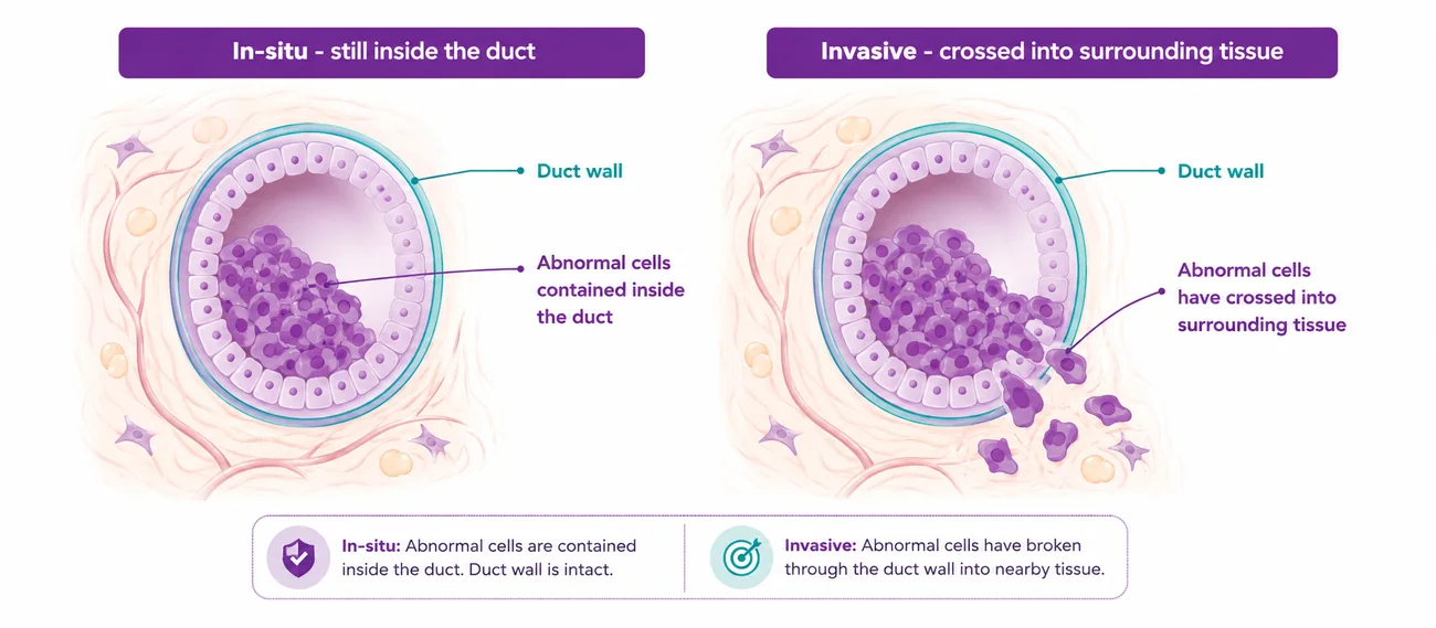
Each of these gets mistaken for something more alarming than it is.
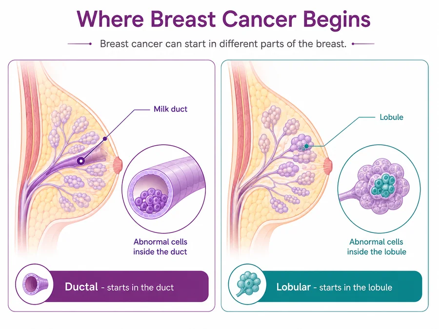
Ductal begins in the tube. Lobular begins in the gland. Neither name means milder or worse.
Like a dairy: one starts in the gland that makes the milk, the other in the pipe that carries it. Same building, different room. Not a severity split — lobular can be harder to spot on scans, so extra imaging is sometimes arranged, but that is detection, not danger.
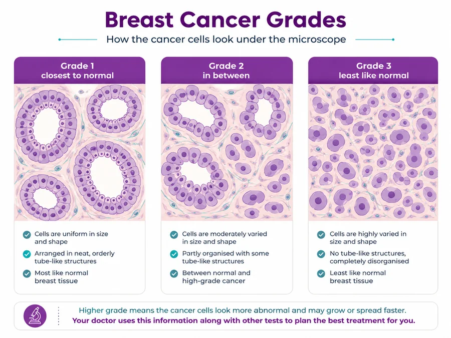
Grade 1 cells are still in neat rows. Grade 3 have stopped resembling normal tissue. Full explanation →
Stage is how far the fire has spread through the building. Grade is how hot it’s burning. Two different questions, answered separately. A higher grade sounds worse, but faster-dividing cells are often the ones treatment acts on most.
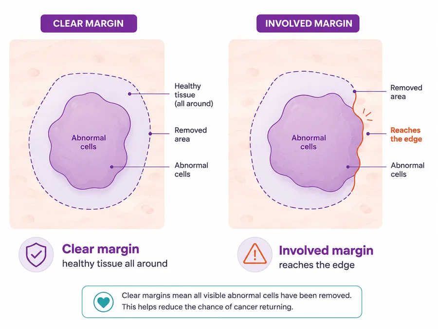
"Clear" means no cancer cells were found at the cut edge. "Involved" means they reached it.
Like cutting a stain out of cloth with a border of clean fabric all the way around, so nothing is left behind. If the margin isn’t clear, a further procedure can be recommended — that is planned for from the start, not a sign the surgeon failed.
Talk to our team
Call the helpline or leave your details, and someone will help you arrange a consultation at the CION centre nearest you. One helpline serves every CION centre.
Together these describe what fuels the cancer, and therefore which treatments can work against it.
The cancer cell has locks on its surface. Hormones in your body are the keys. When a key turns a lock, the cell is told to grow. Blocking treatment either takes the keys away or jams the locks.
HER2 works differently — think extra antennae picking up the “grow” signal far too loudly, with targeted treatment capping the antennae. Keep the two separate in your head: they are different mechanisms and lead to different treatments, which is why the report lists all three.
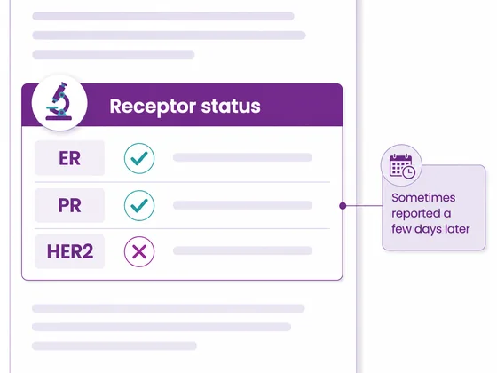
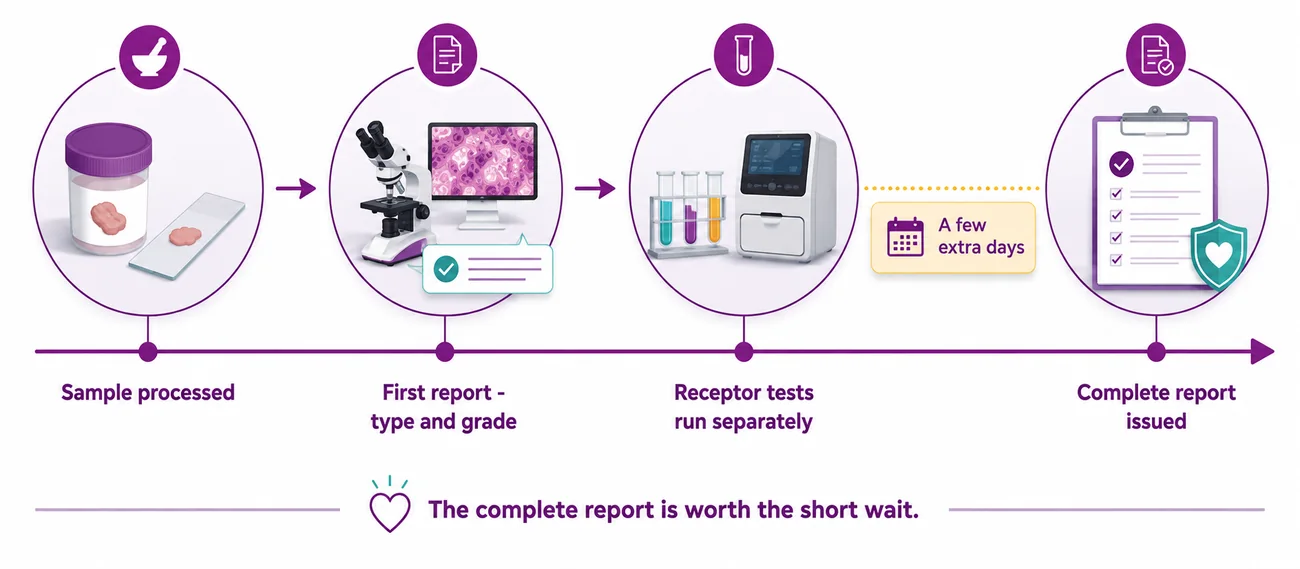
The first report is the quick test. The receptor tests are the detailed one that takes a few more days — like a basic soil result coming back fast while the full mineral breakdown follows. Waiting is not losing time: starting before receptor status is known risks starting the wrong plan, and breast cancer is very rarely a same-week emergency.
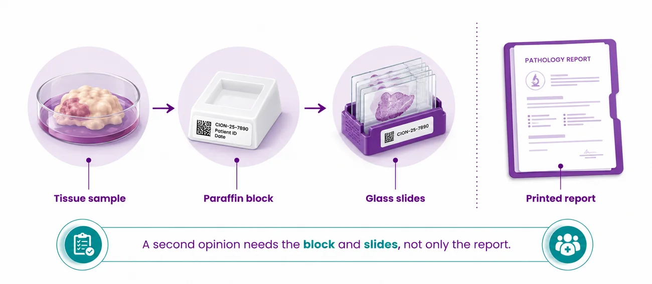
The block and slides are the negative. The printed report is only the description of the photograph. Any second opinion or repeat receptor test needs the negative, not the description. You are generally entitled to request them — ask the laboratory how, keep the receipt, and store them with your reports.
Set the three results as they appear on your report.
The most common combination. The cancer responds to hormone signals, so treatment that blocks those signals is usually part of the plan, often over a number of years. Other treatments may be added depending on stage and grade.
General education, not advice about your case, and not a diagnosis. Your plan depends on stage, grade, general health and your own preferences — set by your oncologist after a multidisciplinary tumour board reviews the case.
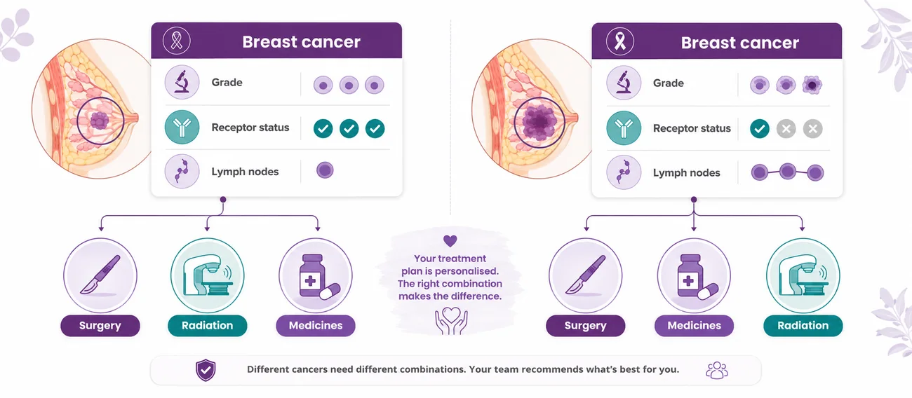
Two women can both be told they have breast cancer and be given completely different plans. That is not a mistake, a cost decision, or one of them getting better care. The plan follows the report.
Two people can both say “my car won’t start” and need completely different repairs. Same complaint, different cause, different fix. Neither plan is the better one — they are individual, never tiered by cost or by how hard anyone is trying.
Comparing plans with a relative or neighbour is one of the most common reasons people lose confidence in their own treatment. It is worth resisting.
Want a second opinion on your plan? Bring your report, your slides and your block. We will tell you honestly whether your current plan is the one we would recommend.
Book a second opinionIf receptor results aren't on your page yet, ask when they're due. Don't plan treatment without them.
The lab holds your tissue. Any second opinion needs it — not just the printout.
Reports, scans, slides, prescriptions, ID and scheme card in a single folder.
Newly diagnosed and unsure what happens now? Your first 7 days, step by step →
Two short explainers covering the same ground as this page. Nothing loads or plays until you press it.
Trained at AIIMS, Tata Memorial, and leading international centres. Combined 150+ years of experience. Every complex case is reviewed by 3+ of them — together.

MBBS(Gold Medal), DNB(General Medicine), DM(Medical Oncology)(Gold Medal)

MBBS, MD(General Medicine), DM(Medical Oncology)(Adyar,Chennai), ECMO, MRCP SCE(UK)

MBBS, MD (General Medicine), DrNB (Medical Oncology), ECMO, MRCP SCE (Medical Oncology) (UK)



MBBS (AIIMS), MS (Surgery) (AIIMS), DNB (Surgical Oncology), MRCS (Edinburgh)



MBBS, MS(General Surgery), M.Ch(Surgical Oncology), FMAS, FARIS(Ongoing)






MBBS, MS (General Surgery), DrNB (Surgical Oncology), FALS Oncology

Want a specific doctor for your case? Mention them when booking.
Book Free ConsultationShare your name and number — we'll call you back within 30 minutes to schedule your consultation.
Where to find us
Addressed by landmark, because that is how this city navigates. Each centre also names the areas it serves, so you can place it without a map. Consultation and day-care Chemotherapy run at every one of them.
Talk to our team
Call the helpline or leave your details, and someone will help you arrange a consultation at the CION centre nearest you. One helpline serves every CION centre.
The six we hear most often after a biopsy result. Tap any question to open the answer.
This belief is widespread and not supported by evidence. A biopsy is a standard, controlled procedure done by trained specialists, and it does not cause breast cancer to spread. The real risk runs the other way — avoiding or postponing a biopsy delays diagnosis and gives a treatable cancer more time to grow. If a family member has raised this, bring them to the consultation so the team can explain how it is done.
The first report covers type and grade; the receptor tests run afterwards and add a few days. Breast cancer is very rarely a same-week emergency, and starting before the complete picture is known risks starting the wrong plan. A short wait for complete information is generally the safer choice, not the riskier one. If the wait stretches further, it is usually because a borderline result has been sent for a confirming test, not because anything has been forgotten. Ask when the complete report is expected, and use the time to gather your scans and earlier reports into one file.
Grade 3 means the cells look least like normal breast tissue and appear to be dividing quickly. But faster-dividing cells are often the ones most affected by treatments that act on dividing cells. It is not the same as stage. Grade is decided by scoring three separate features of the cells and adding them together, so it is a reasoned total rather than one pathologist’s impression. Your oncologist reads it alongside the stage, the receptor status and your general health rather than reacting to any single line on the page.
The laboratory keeps the tissue as a paraffin block with prepared slides, and you are generally entitled to request them. Any second opinion, pathology review or repeat receptor test needs the original tissue rather than just the printed report. Patients who arrive without them frequently lose several weeks retrieving them. Ask how to request them, keep the receipt, and store them with your reports. It costs nothing to have them ready, and it can save several weeks if you later decide you want another opinion.
Keeping it hidden tends to isolate the patient and delay decisions. Cancer is a disease with biological causes — not a punishment, not a curse, and nothing anyone deserves. Families told early can help with appointments, travel from the districts, scheme paperwork and the day-to-day load. Many patients find the fear of telling people was worse than telling them. If you are worried about how one particular relative will react, tell someone you trust first and let them help you with the rest.
Traditional practices may support comfort and wellbeing, but using them instead of evidence-based treatment is one of the most dangerous choices available, because it spends the window when the cancer is most treatable. Anything complementary should be discussed with the oncology team rather than substituted for treatment. Do tell your oncologist what you are taking, including supplements and traditional preparations, because some can interact with treatment or affect your blood results. Nobody will judge you for asking.
Still have a question this page didn't answer? Ask it directly — we would rather you called than guessed.
Talk to an oncologistUnderstanding the report is the first step. At CION these run alongside treatment, not as afterthoughts.
Appetite, weight and strength through treatment.
Body image and identity support, patient and family.
Hereditary risk for you and close relatives.
Comfort and symptom support at any stage.
Dedicated pain control as part of the plan.
Shoulder mobility and lymphoedema care.
An oncologist will read your report and tell you what it means for you — including whether a second opinion is worth getting. Bring the printout, and the slides and block if you have them.
Toll free 1800 202 8726 · Advancing Care. Passionately.