Being referred for a breast biopsy is not a cancer diagnosis. Across breast clinics, roughly 75 to 80 percent of all breast biopsies turn out to be benign — non-cancerous. A biopsy exists to settle uncertainty: for most women it ends in reassurance, and for the minority who do have cancer, it enables an early, accurate diagnosis — the single biggest advantage for successful treatment. Source: NCCN guidelines / peer-reviewed breast-clinic data.
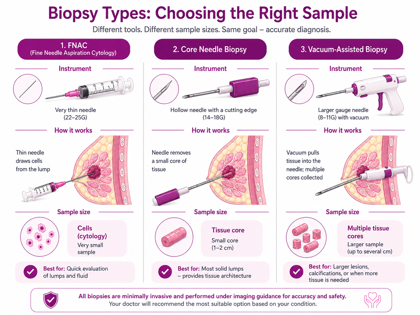
There is no single "breast biopsy" — the right method depends on the size, location and nature of the finding. Your specialist chooses the least invasive technique that will still give a reliable answer. These are the main types, from the simplest needle test to a surgical biopsy.
The preferred method for a breast lump. A hollow needle removes one or more small cylinders (cores) of tissue, usually under ultrasound guidance and local anaesthetic. Because it samples actual tissue — not just cells — it can tell invasive from non-invasive cancer and allows ER, PR and HER2 testing.
A gentle vacuum draws tissue into the needle, so several samples are collected through a single small insertion. It removes more tissue than a standard core needle, which improves accuracy for tricky targets like microcalcifications, and is often done under stereotactic (mammographic) or ultrasound guidance.
A very thin needle draws out cells (not a tissue core) for cytology. It is quick, low-cost and useful for draining a cyst or sampling an enlarged lymph node, but it cannot tell invasive from in-situ cancer — so for a solid breast lump a core needle biopsy is usually preferred over FNAC.
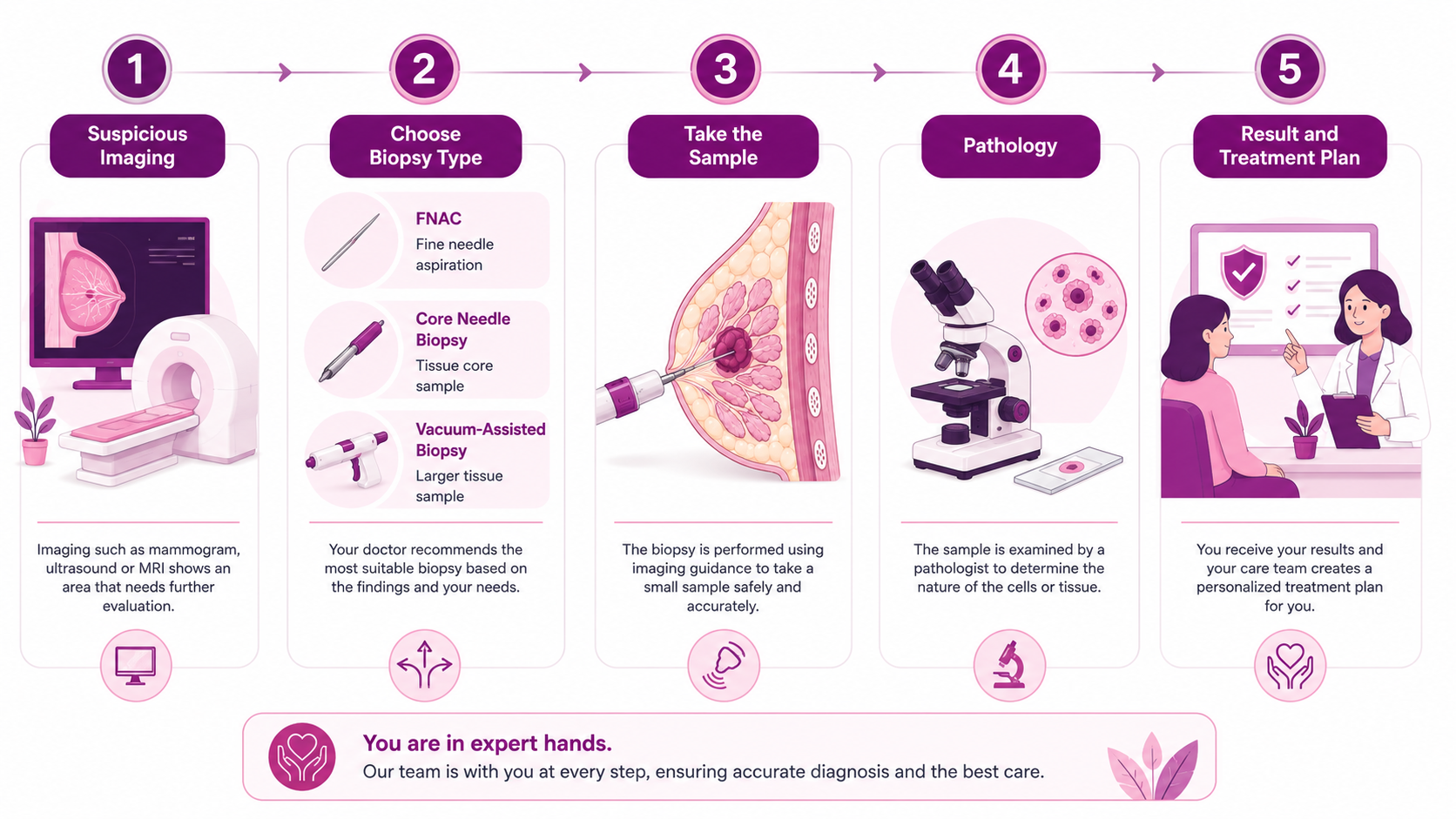
A core needle biopsy is a same-day procedure — there is no general anaesthesia, no overnight stay, and most people are back to normal the next day. The needle insertion itself takes only a minute or two; the whole appointment, including imaging, usually runs 30 to 60 minutes. Here is exactly what happens, step by step.
You lie down and the radiologist uses ultrasound (or a mammogram for stereotactic biopsy) to locate the exact spot to be sampled and to guide the needle in real time.
The skin and breast tissue over the target are numbed with a local anaesthetic injection. You stay fully awake, but the area is numb — you should feel pressure, not pain.
A tiny nick is made in the skin and the biopsy needle is guided to the lesion. Several small cores of tissue are taken. You may hear a soft click or snapping sound from the spring-loaded device — this is normal.
A tiny, painless titanium marker clip may be placed at the site so the exact spot can be found again later if treatment or further imaging is needed. It is harmless and stays in place.
Pressure is applied to stop any bleeding, the small wound is closed with a steri-strip or small dressing (no stitches needed), and you rest briefly before going home. You can usually drive yourself if only local anaesthetic was used.
We're never more than 30 minutes away. Same panel of specialists at every centre. Same tumour board reviews. Same NCCN protocols. Pick the closest one and call directly — or let us pick for you.
Not sure which centre fits best? Tell us where you are — we'll suggest the closest one with the right specialists.
Help me pick the right centreTravelling for treatment? We may have a centre right where you are.
Don't see your city? Call 18002028726 — we'll find your nearest CION partner centre.
Surgical, medical and radiation oncologists review every case together in a multidisciplinary tumor board — part of 17 senior specialists across CION.


MBBS(Gold Medal), DNB(General Medicine), DM(Medical Oncology)(Gold Medal)

MBBS, MD(General Medicine), DM(Medical Oncology)(Adyar,Chennai), ECMO, MRCP SCE(UK)

MBBS, MD (General Medicine), DrNB (Medical Oncology), ECMO, MRCP SCE (Medical Oncology) (UK)


MBBS (AIIMS), MS (Surgery) (AIIMS), DNB (Surgical Oncology), MRCS (Edinburgh)


MBBS, MS(General Surgery), M.Ch(Surgical Oncology), FMAS, FARIS(Ongoing)


MBBS, MS (General Surgery), DrNB (Surgical Oncology), FALS Oncology

Want a specific doctor for your case? Mention them when booking.
Book Free ConsultationShare your name and number — we'll call you back within 30 minutes to schedule your consultation.
Confidential, doctor-led, and free for your first consultation. Call us on 1800-202-8726 or request a callback - we'll guide your next step.
There is no single price for a biopsy — it depends mainly on the technique and the laboratory testing required. The table below is an indicative guide for common biopsies; your exact, confirmed price is shared once your prescription and reports are reviewed.
| Biopsy type | Starting price |
|---|---|
| FNAC (fine needle aspiration) | From ₹8,000 |
| Core needle biopsy | From ₹7,500 |
| Image-guided biopsy (ultrasound / CT) | ₹7,000–13,000 |
| Bone marrow aspiration & biopsy | From ₹12,000 |
| Endoscopic biopsy | By referral |
| Surgical (excisional / incisional) biopsy | ₹20,000–25,000 |
| Breast biopsy | From ₹6,000 |
| Prostate biopsy | From ₹12,000 |
| Liquid biopsy (blood test) | ₹35,000–37,000 |
Prices shown are indicative and may vary by site, image guidance and the pathology testing required. Histopathology and any special tests (IHC / molecular) are charged separately unless stated.
A clear, up-front cost for the biopsy and the pathology — no hidden add-ons, no tests you don't need.
Concessional diagnostic pricing for cancer patients, with expert-reviewed pathology reporting.
Cashless private insurance and EMI support available — our team helps you with the paperwork.
Your first consultation is free, so you can understand your options and costs before deciding.
A panel of 17 super-specialist oncologists across medical, surgical and radiation oncology.
Complex cases reviewed by 3+ specialists together — not one doctor's opinion.
A trusted network across Telangana and AP, rated 4.8/5 by patients on Google.
No unnecessary tests, transparent costs, and a free first consultation for all cancer patients.
*1-year survival. Source: ICMR / National Cancer Registry Programme (NCRP), compared with CION patient outcomes. CION figures are network outcomes; national figures are population averages and do not predict an individual's result.

Been advised a breast biopsy? In a free, 45-minute consultation we review your scan or report, explain what it means in plain language, and recommend only the biopsy you actually need — nothing more. Every result is reviewed by our tumor board, so decisions are made by a team, not a single opinion.
No, a needle breast biopsy is not painful. The area is numbed with a local anaesthetic injection, so during the procedure most people feel pressure rather than pain. You stay awake and can tell the radiologist if you feel any sharp sensation, in which case more anaesthetic is given. Afterwards there may be mild soreness and some bruising for a few days, which is easily managed with simple painkillers like paracetamol. A surgical (excisional) biopsy involves a small cut and may use sedation, but it too is designed to keep you comfortable throughout.
For a routine breast biopsy, the pathology report is usually ready within about 3 to 7 days, and sometimes sooner. The tissue has to be carefully processed, stained and examined by a pathologist, which cannot be safely rushed. If special tests are needed — such as ER, PR and HER2 immunohistochemistry, or FISH and molecular testing on a confirmed cancer — these add a few more days. At CION, your report is reviewed by our tumor board and explained to you by a specialist rather than simply handed over, and anything urgent is fast-tracked.
A core needle biopsy (CNB) uses a hollow needle to remove small cylinders of actual tissue, while FNAC (fine needle aspiration cytology) uses a very thin needle to draw out individual cells. The key difference is that a core needle biopsy samples intact tissue, so it can tell invasive cancer from non-invasive (in-situ) disease and allows ER, PR and HER2 testing. FNAC is quicker and cheaper and is useful for draining a cyst or checking a lymph node, but for diagnosing a solid breast lump, a core needle biopsy is usually preferred because it gives more complete and reliable information.
No. This is a very common worry, but it is not supported by medical evidence. Modern biopsy techniques are safe and do not cause cancer to spread through the body. In fact, delaying or refusing a recommended biopsy is far riskier, because it can delay a diagnosis and the start of effective treatment. A biopsy simply removes a tiny sample so doctors can identify exactly what they are dealing with. The benefit of an accurate, early diagnosis vastly outweighs this unfounded fear.
In Hyderabad, a breast biopsy typically costs between Rs. 3,500 and Rs. 15,000, depending on the type of biopsy, whether image guidance is used, and the pathology testing required. A core needle biopsy is usually at the lower end, while vacuum-assisted and surgical biopsies cost more. The histopathology (lab analysis) is generally charged separately, and special tests such as ER/PR/HER2 add to the total. At CION we keep costs transparent and offer up to 50 percent off diagnostics for cancer patients, with expert-reviewed reports. Your first consultation is free — request a callback for an exact estimate.
The report first states whether the tissue is benign (non-cancerous) or malignant. Most are benign. If cancer is found, the report describes whether it is in-situ or invasive, the histologic type (such as invasive ductal carcinoma), the grade (1 to 3, how aggressive it looks), and the receptor status — ER, PR and HER2 — which decides whether hormone therapy or targeted anti-HER2 therapy will help. It may also include a Ki-67 score showing how fast cells are dividing. These details together guide your personalised treatment plan, which is why our team explains every part of the report to you.
Yes, a needle breast biopsy is a day-care procedure. It is done under local anaesthetic, takes only 15 to 20 minutes for the needle part, and you go home the same day with no overnight stay. If only local anaesthetic was used, you can usually drive yourself home. If you are given sedation — more common with a surgical biopsy — you should arrange for someone to drive you. It is sensible to avoid strenuous activity and heavy lifting for about 48 hours afterwards to limit bruising, but most people return to normal life the same or next day.
Preparation is usually simple. For a needle biopsy under local anaesthetic you do not normally need to fast. Tell your doctor about all medicines you take, especially blood thinners such as aspirin or warfarin, as you may be advised to pause them for a few days beforehand. On the day, avoid applying deodorant, powder, lotion or perfume to the breast and underarm, and wear a comfortable two-piece outfit and a supportive bra. If sedation or general anaesthesia is planned for a surgical biopsy, you will be told how long to fast. Your CION team will give you exact instructions for your specific procedure.
No. Being referred for a breast biopsy does not mean you have cancer. It simply means a finding needs to be checked to be certain. In fact, around 75 to 80 percent of all breast biopsies turn out to be benign — that is, non-cancerous. Doctors recommend a biopsy because imaging and examination alone cannot give a definite answer, and the only responsible step is to confirm one way or the other. For most women, a biopsy ends in reassurance. For the minority who do have cancer, an early, accurate diagnosis is the single best advantage for successful treatment.
Browse our complete guide to breast cancer — types, symptoms, tests and treatments. Tap any topic to read more.